")
")
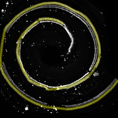
The cochlea is the auditory organ of the inner ear, containing inner and outer sensory hair cells, organized in respectively one and three rows inside the organ of Corti. The cochlear spiral structure with tonotopic, sensorineural organization makes precise histological studies demanding, however the information provided supports and facilitates interpretation of functional audiometric studies.
"We aim to develop automated algorithms for quantifying cochlear hair cells in sections of 200 μm from the cochlear apex to base to facilitate higher throughput histology for preclinical drug development. Similar approaches exist for other tissues, but none are applicable to the particular organization of the cochlea."
Temporal bones of adult Wistar rats were extracted following PFA fixation and the organ of Corti dissected after decalcification (EDTA 10%). Hair cells were immunolabelled with Myosin VIIA (1:1000)/Alexa Fluor 594 (1:700) conjugated donkey anti rabbit. 3D image stacks of the organ of Corti were obtained using a Zeiss Apotome Fluorescence microscope and the entire structure was reconstructed as a mosaic of 4 stacks.
Sensorion asked MRI to find a solution for the image analysis problem. We developed a protocol based on Imaris and ImageJ.
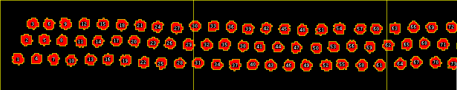
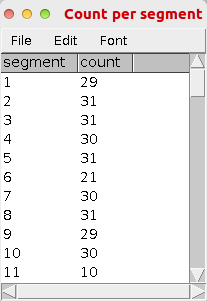
From the stitched 3D stack a binary mask of the inner and outer hair cells was created using the spot detection algorithm of Imaris. The mask image stack was imported into ImageJ. A maximum intensity projection was applied. The user selected cochlea was then straightened. On the straightened cochlea the cells are automatically counted for each segment of 200μm using an ImageJ toolset.

In a first time we presented the work at the 53rd Inner Ear Workshop in Montpellier:
Saleur, A., Baecker, V., Dyhrfjeld-Johnsen, J., poster: AUTOMATED CELL COUNTING IN COCHLEAR HISTOLOGICAL SAMPLES, "53. Workshop Inner Ear Biology, Montpellier, 2016
Sensorion continued to use the tools developed by MRI in its research to find a biomarker for hearing loss, resulting in the paper:
Parham, K., Sohal, M., Petremann, M., Romanet, C., Broussy, A., Tran Van Ba, C., and Dyhrfjeld-Johnsen, J. (2019). Noise-induced trauma produces a temporal pattern of change in blood levels of the outer hair cell biomarker prestin. Hearing Research 371, 98–104.
"The paper, published in the peer-reviewed monthly journal Hearing Research, found that the outer-hair-cell-specific protein prestin, in circulation, may act as a biomarker for sensory hair cell damage and death. Prestin levels in circulation change after acoustic trauma in a preclinical model of sudden sensorineural hearing loss and the pattern of change is dependent on the severity of injury, with an early rise in prestin levels after noise exposure potentially predictive of permanent hearing loss."
“Thus, an outer hair cell (OHC)-specific circulating biomarker may have both research and clinical applications, such as development of therapeutics and early diagnosis,” concluded Dr. Kourosh Parham, associate Professor and Director of Research in UConn Health’s Division of Otolaryngology, Head & Neck Surgery. “These results suggest that there is a temporal pattern of change in serum prestin levels after acute hearing loss that is related to severity of hearing loss. Circulating levels of prestin may be able to act as a surrogate biomarker for hearing loss involving OHC loss.” (businesswire)