images/analyse-de-donnees/window-2-fill_3.png

Software

The facility provides access to proprietary analysis software, open source software and tools developed in house. We also provide access to deep-learning solutions including the training on your own data on our machines.

proprietary     open-source     deep-learning     tools

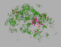
3D Nuclei Clustering Tool

Analyze the clustering behavior of nuclei in 3D images. The centers of the nuclei are detected. The nuclei are filtered by the presence of a signal in a different channel. They clustered with the density based algorithm DBSCAN. The nearest neighbor distances between all nuclei and those outside and inside of the clusters are calculated.

Download and further informations: 3D_Nuclei_Clustering_Tool on github

Adipocytes Tools

Adipocytes, are the cells that primarily compose adipose tissue, specialized in storing energy as fat. The tool allows to segment the adipocytes in images from histological sections. Once segmented the size of the cells can me measured and their morphology can be analyzed.

Download and further informations: Adipocytes Tools sur github

Arabidopsis Seedlings Tool

The Arabidopsis Seedlings Tool allows to measure the surface of green pixels per well in images containing multiple wells. It can be run in batch mode on a series of images. It writes a spreadsheet file with the measured area per well and saves a control image showing the green surface that has been detected per well.

Download and further informations: Arabidopsis Seedlings Tool on github

Cell profiler

Software for quantitative image analysis and task automation. No programming skills are required, the software works via the creation of image analysis pipelines.

Cellpose

Cellpose allows to segment cells of different types from different imaging modalities. The deep-learning network is pre-trained with images of 70000 cells. Cellpose uses a vector flow representation of cells. The input is  an image of the cells and optionally an image of stained nuclei.

MRI added a batch processing button, which allows to run the batch segmentation of cells from the graphical user interface instead of using the python from the command-line.

See also:

  • Cellpose (Presentation for the MRI image analysis group,  05.02.2020)
  • Cellpose reloaded (Presentation for the MRI image analysis group, 04.03.2021)

FIJI / ImageJ

2D and 3D image processing and analysis software. Extensible via macros (IJ-macro language), scripts (python, javascript, ...)  and plugins (java).

  • Available on all workstations of the facility.
  • ImageJ/Fiji