images/analyse-de-donnees/trophy-line_10.png

This page lists some examples of image analysis projects that have been successfully realized  by MRI in the past.

Automated and multiparametric image analysis on osteoclast cells

Automated and multiparametric image analysis on osteoclast cells

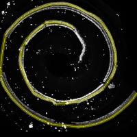
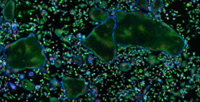
The Anne Blangy team (CRBM) wanted to perform a screening to identify genes involved in the cellular differentiation of mature osteoclasts using siRNA. For this purpose, mouse femur cells were cultured and differentiated into osteoclasts in 24-well microplates (Figure A). The ArrayScan/Cellomics was used for automated image acquisitions as shown in Figure B. The objective is to be able to segment, count, measure the size and differentiation stage of osteoclasts, in order to evaluate the effect of the tested siRNAs.

Continue Reading

Automated cell counting in cochlear histological samples

The cochlea is the auditory organ of the inner ear, containing inner and outer sensory hair cells, organized in respectively one and three rows inside the organ of Corti. The cochlear spiral structure with tonotopic, sensorineural organization makes precise histological studies demanding, however the information provided supports and facilitates interpretation of functional audiometric studies.

"We aim to develop automated algorithms for quantifying cochlear hair cells in sections of 200 μm from the cochlear apex to base to facilitate higher throughput histology for preclinical drug development. Similar approaches exist for other tissues, but none are applicable to the particular organization of the cochlea."

Continue Reading