")
")
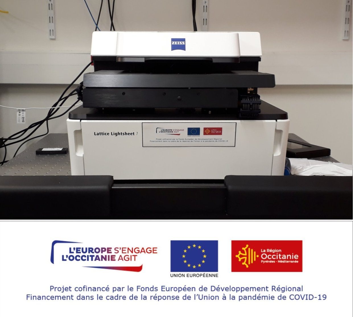
Lattice Light Sheet
Tarif établissement partenaire: 15.66 €/heure
Tarif établissement public et privé : 55.41 €/heure
Brève description :
The Zeiss Lattice Light Sheet Microscope 7 is a light sheet microscope with structured light for 3D imaging of live and fluorescent sample with a maximum of 100µm thickness. The 3D acquisitions are fast and induce less photobleaching and photoxicity thanks to the excitation in single plane.
Objectives:
-excitation equivalent to 13.3X 0.4 NA
-emission equivalent to 44.8X 1.0 NA with a free-form phase lens to correct the thickness of the coverslip
-miniscus lens with water immersion between the emission objective and the coverslip of the sample.
Detector :
1 sCMOS Hamamatsu ORCA Fusion camera
(2048*2048, 6.5µm pixel size)
Mode of acquisitions:
lsequential multi-color light sheet imaging, X-stacks, multi-positions, mosaics, multi-time points.
Controled by the Zen blue 3.7 software.
Processing of images by deconvolution, deskew, cover glass transformation.
lsequential multi-color light sheet imaging, X-stacks, multi-positions, mosaics, multi-time points.
Controled by the Zen blue 3.7 software.
Processing of images by deconvolution, deskew, cover glass transformation.
Excitation sources:
diode 488nm
diode 561nm
diode 640nm
5 light sheet structured by a SLM (Spatial Light Modulator) with variable lengh x thickness :
15µm x 550nm; 15µm x 650nm; 30µm x 700nm; 30µm x 1000nm; 100µm x 1400nm; 100µm x 1800nm.
Spectral selection:
BP 570-620 + LP 655
BP 495-550 + LP655
diode 488nm
diode 561nm
diode 640nm
5 light sheet structured by a SLM (Spatial Light Modulator) with variable lengh x thickness :
15µm x 550nm; 15µm x 650nm; 30µm x 700nm; 30µm x 1000nm; 100µm x 1400nm; 100µm x 1800nm.
Spectral selection:
BP 570-620 + LP 655
BP 495-550 + LP655
Sample Support:
the sample is preparaed as usual for inverted microscope, the sample support has to have a bottom glass 0.17mm thick.
The motorized stage can have specific inserts for 35mm dish, slide and multi-well labtek.
Incubation:
tempretaure from RT to 40°C
CO2 from 0 to 5%
the sample is preparaed as usual for inverted microscope, the sample support has to have a bottom glass 0.17mm thick.
The motorized stage can have specific inserts for 35mm dish, slide and multi-well labtek.
Incubation:
tempretaure from RT to 40°C
CO2 from 0 to 5%