English

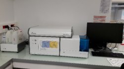
NOVOCYTE ACEA 2 (violet)


Partner lab price : 21.78 €/hour
Public and private lab price: 59.44 €/hour
Brief description:
Flow cytometry cell analyzer - ACEA
3 lasers, 2 physical parameters and 13 fluorescences
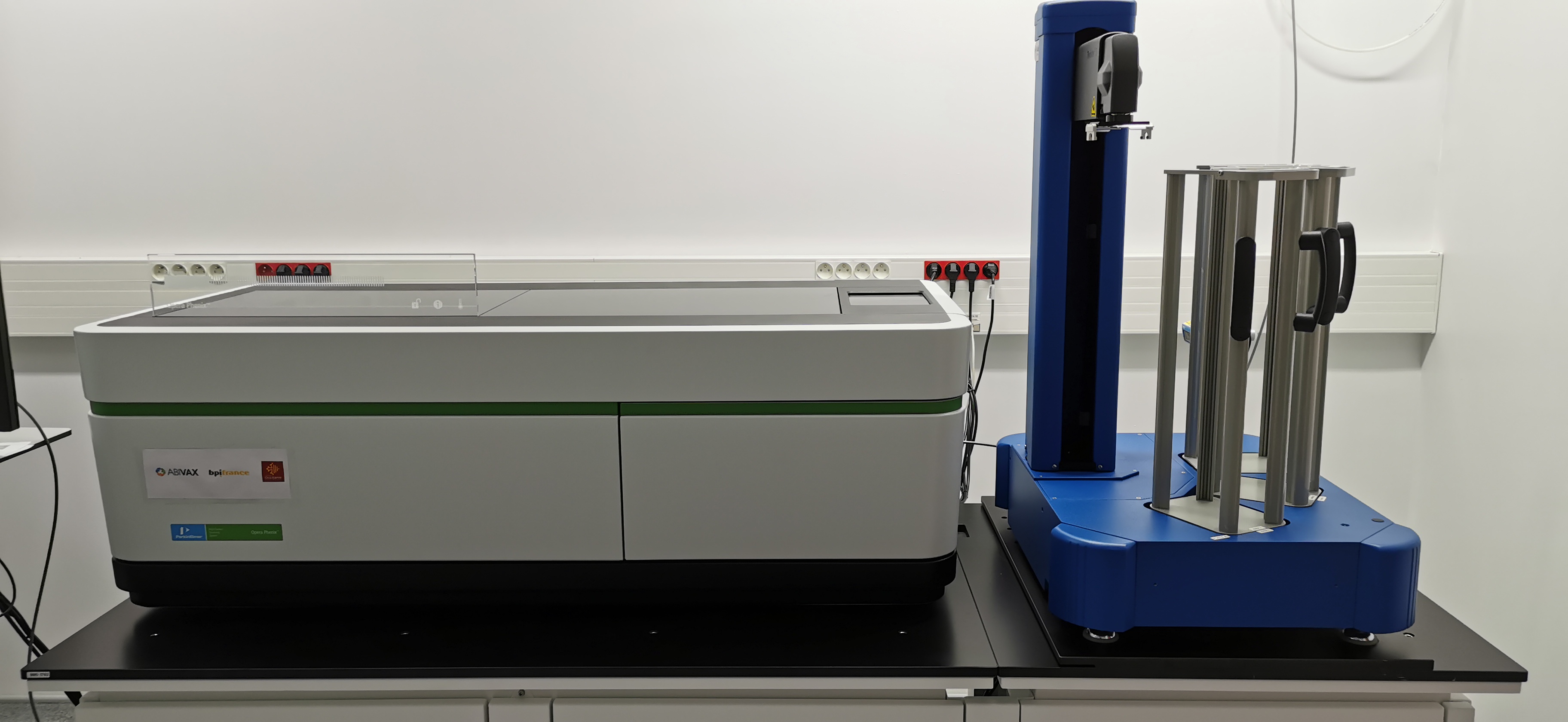
Plate and tubes loader
Controlled by the NovoExpress software (ACEA)
Blue laser excitation- 488nm :
filter 530/30 BP: FITC, Alexa Fluor 488, GFP, CFSE, Sytox Green, FAM-Azide, YFP, BB515
filter 572/28 BP : PE, (dTomato)
filter 615/20 BP : PE-TexasRed, PI, (mcherry), PE-CF594
filter 675/30 BP : PerCp, PerCp-cy5.5, PerCp-eF710, 7AAD, PE-Cy5, PE-cy5.5
filter 780/60 BP : PE-Cy7
Red laser excitation 640 nm:
filter 675/30 BP : APC, A647, Crimson, Cy5, Sytox Red, eF660
filter 780/60BP : APC-Cy7, APC-H7, APC-AF750, APC-eF780
Violet laser excitation 405 nm:
filter 445/45 BP : BD Horizon V450, Pacific Blue, AF405, BV421, Sytox blue, Dapi, BFP
filter 530/30 BP : BD Horizon V500, AmCyan, BV510
filter 572/28 BP : Pacific Orange, BV570
filter 615/20 BP : BV605
filter 675/30 BP : BV650
filter 780/60 BP : BV785, (BV750, BV711)

Acquisition of 30 000 events/second

Intracellular or cell-surface staining, fixed or alive cells

L2 laboratory completely equiped : hood, centrifuge, pipets
Ideal operating temperatures between 19 and 26°C

Open-Desk Sessions

Do you have questions about image analysis? Do you need help with a script or macro? Are you planning to start an image analysis project with us? Then come join us at the next open-desk session. The access is free and no inscription is needed, but we ask you to kindly give us some information, that will help us planning the sessions.

The upcoming sessions are (please click on a date to go to the inscription):


You need a QR-code and ID to enter the campus. You can demand the creation of a QR-code here. You need to select CRBM-MRI as the  laboratory you want to visit and "Volker Baecker" as approver. Should you not have access, please ask for the qr-code via email at This email address is being protected from spambots. You need JavaScript enabled to view it..

The IGMM is the building next to the CRBM.

Opera Phenix


Partner lab price : 37.04 €/hour
Public and private lab price: 101.89 €/hour
Associated technologies:
  • High Content Screening
  • Image analysis

  • Brief description:
    Automated epifluorescent widefield and confocal (spinning disk) microscope with 2 cameras. Ideal for high throughput measure and analysis of cellular content with a large number of samples (cellules, sphéroïdes...) fixed or live and in microplate format.
    Piloted by Harmony software
    Objectives:
    5X air 0.16NA Plan Apochromat
    10X air 0.3NA EC Plan Neofluar
    20X air O.4NA LD Plan Neofluar
    20X air O.8NA Plan Apochromat
    63X eau 1.15NA LD C-Apochromat

    Twister III robotic arm to automatically charge the microplates
    Excitation Source:
    4 Lasers : 405 nm, 488 nm, 561 nm et 640 nm

    Emission filters:
    435-550 nm, -435-480 nm, 500-550 nm, 570-630 nm et 650-760 nm

    Can read any format of microplate (6 à 1536 wells)
    Detector:
    2 cameras sCMOS 4.7Mpx 16bits
    (2160 x 2160 pixels, 6.5µm pixel size)

    Environmental control: temperature and CO²
    Applications:
    -multidimensional automated acquisition: YXZ, multi-channels, multi-fileds, multi-wells, multi-plates, timelaps
    -on-the-fly image analysis: personnalized protocol based on different building blocks pipeline
    -Preciscan XYZ: detection of rare objects or events at low magnification and automated ReScan at higher magnification
    -Analysis and classification of objects by Machine Learning

    Optogenetics

    
    Tarif établissement partenaire: 15.66 €/heure
    Tarif établissement public et privé : 55.41 €/heure

    Brève description :
    Epifluorescence Nikon Ti2 inverted microscope for the 2D and 3D videomicroscopies coupled to the multipoint photomanipulation thanks to the DMD module (digitale micromirror device).
    Controlled by the NIS Element software
    Objectives:
    10X Plan Apochromat 0.45NA air
    20X Plan Apochromat 0.75NA air
    40X Plan Fluor 1.3NA oil
    60X Plan Apochromat 1.4NA oil
    100X Plan Apochromat 1.4NA oil

    Detector:
    1 CMOS back-illuminated Prime95B Photometrics camera
    (1200*1200, 11µm pixel size)

    Incubation:
    control of temperature and CO2

    Illumination Sources:
    1 Lumencor system with 6 LED and excitation filters for:
    DAPI, CFP, GFP, YFP, Cy3, Cherry, Cy5
    1 Luminencor system avec 6 LED and excitation filters for the DMD module:
    DAPI, CFP, GFP, YFP, Cherry

    Emission Filters
    DAPI, CFP, GFP, YFP, Cy3, Cherry, Cy5


    Types of acquisition with the DMD module:
    -point photoactivation, with pre-defined regions or on the fly in simultaneous multiple points or regions, particularly adapted for the optogenetics experiments.

    Definition of the number, size, form of the areas in NIS Elements software.

    Compatible sample carriers:
    35mm diameter dish with a bottom glass (#1.5), glass slide with a glass coverslip (#1.5) or multi-well plate with a bottom glass (#1.5)
    

    Our facilities / softwares

    Other software applications are listed on the website biii.info

    Facility / Softwares2D analysis3D analysisObject
    Segmentation
    AutomationSignal
    processing
    Easy to useDownloadable on WebPaying on
    analysis station
    ImageJ  +++  +  +++  +++    +++  +  
    Fiji  +++  +  +++  +++    +++  +  
    Metamorph  ++    ++  ++    +++    +
    Icy  +++ +  +++  ++    +++  +  
    Zen  ++        ++  +++    +
    LAS-AF  ++  +      +  +++    +
    Definiens                +
    Imaris +  +++  +++  +    ++    +
    Volocity  +  +++        +++    +
    ISE-MESH Tools    +++        + +  
    Avizo    +++  +++      +    +
    Drishti    +++            
    Studio  +++    ++  +++    +    +
    Cell Profiler  +++    +++  +++    +  +  
    Matlab  +++  +++  +++  +++    +  +  +
    Becker&Hickl          +++  ++    +
    Symphotime          +++  ++    +
    eTomo   *          +  

    * Reconstruction of tomogram MET

    Our services


    The MRI facility offers three types of services: the provision of equipment in autonomous manner, the provision of services and collaborative projects. After registering with an MRI engineer and receiving training, any user can use the platform's various equipment after purchasing reference hours. The rates per machine are indicated on their corresponding page

    Purchase orders must specify the name and e-mail address of the user and the group to which the hours will be assigned.

    They should be sent to This email address is being protected from spambots. You need JavaScript enabled to view it.

    To check your credit hours: log on to the    https://cfms.mri.cnrs.fr/
    and then on the "Reservation" button on the top right. You will then be able to see the balance of all the accounts to which your login is associated

    You're a company

    To use our facilities, you could contact us by phone at +33 (0)6 21 75 76 07 or by e-mail : This email address is being protected from spambots. You need JavaScript enabled to view it.
    You could also have a look at  this flyer.

    TRAINING

    The MRI platform offers throughout the year permanent training courses recognized by our main tutelles and doctoral schools. They are listed in a portfolio of BioCampus workshops   (have a look on BioCampus website).

    Our offer evolves every year :

    - Microscopie à épifluorescence et microscopie confocale: de la base à la pratique

    - Imagerie Avancée: Super-Résolution en imagerie photonique

    - Les bases du logiciel ImageJ

    - Programmation de macros avec ImageJ

    - L’apprentissage automatique « machine learning » appliqué à l’analyse d’images biologiques

    - Analyse d'images 3D

    - Les bases du logiciel OMERO

    - Les bases du logiciel Flow Jo

    Expertise / Collaborative project

    On request, with MRI engineers

    R & D

    On request, with MRI engineers


    Our technologies

    Imagerie de l'os et extraction des structures internes d'intérêt


    Analyses morphologiques comparatives par Géométrie Morphométrique


    Création d'atlas anatomiques virtuels


    Reconstruction de fossiles (rétro-déformation, repositionnement des structures déplacées etc…)


    Our tools

    REGISTER AND BOOK EQUIPMENTS

    To get access to the facility workstations and on-line services, you need to own a MRI account and a login/password.
    To open an account, click on subscription.
    Upon receipt of your inscription your account will be created but inactive.
    You will receive an email inviting you to make an appointment with one of the engineers listed in order to have your registration interview
    If you do not receive any confirmation email within 3 days, please contact the facility administrator (This email address is being protected from spambots. You need JavaScript enabled to view it.).

    If you had lost or forgotten your password please use this link : forgotten password

    Sorting Forms.

    Access to certain cytometry systems requires the completion of a sorting form.
    Sorting form (EN) - Sorting form (FR)

    TRANSFER  STORE  SHARE  DATA

    sFTP
    MRI offer an sFTP file tranfer service, accessible from all of our facilities. In order to use this service you had to have an MRI account and follow the process available below.
    Tutorial

    sFTPJobs
    SftpJobs is a program that allows an intelligent transfer of your data on an Sftp Server. It proceeds and automates the transfer files in background task, even if the user is no longer connected
    Tutorial

     
    OMERO

    OMERO is client-server software for visualization, management and analysis of biological microscope images. 
    From the microscope to publication, OMERO handles all your images in a secure central repository. You can view, organize, analyze and share your data from anywhere you have internet access. Work with your images from a desktop app (Windows, Mac or Linux), from the web or from 3rd party software. Over 140 image file formats supported, including all major microscope formats.


    At MRI we provide access to an OMERO server at https://omero.mri.cnrs.fr.
    You can log in using your MRI credentials and start using it.
    If you want to install the OMERO desktop app, follow the instructions we provide at 
     
    Help pages are accessible at