Partner lab price : 5.36 €/hour
Public and private lab price: 22.32 €/hour
Objectives:
10X UPFLN 0.3NA PH1
20X LUCPLFLN 0.45NA PH1
20X LUCPLFLN 0.45NA RC2
40X LUCPLFLN 0.6NA PH2
40X LUCPLFLN 0.6NA RC2
Detector:
1 ZYLA 4.2 MP scMOS camera
(2048*2048 pixels, 6.5µm pixel size)
Incubation:
temperature and CO2 control
Type of acquisition :
-widefield epifluorescent microscopy
-transmitted light microscope in bright field, phase contrats or DIC-like mode.
The Multi-dimensional Acquisition module in Metamorph controls the multi-series of images in multi-channels, multi-positions, multi-Z planes, multi-timepoints.
Filter cubes:
Hoechst
FITC
Cherry
Cy5
Illumination sources:
White LED Xcite
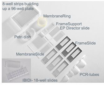
Compatible sample carriers:
multi-well plastic plate; slide or bottom glass/plastic labtek, 35mm diameter disk bottom glass/plastic
Continue Reading
Posted in Videomicroscopy
Partner lab price : 5.36 €/hour
Public and private lab price: 22.32 €/hour
Objectives :
Zeiss 2.5X/0.075 EC-Plan-Neofluar
Zeiss 10X/0.25 A-Plan Ph1
Zeiss 20X/0.4 Plan Neofluar bague Korr
Zeiss 40X/1.3 Plan-Neofluar oil
Zeiss 63X/1.4 PL-APO oil
Zeiss 100X/1.4 Ph3 Plan Apo oil
Fluorescence filters cubes :
- DAPI ex G365, FT395, em BP445/50
- GFP ex BP475/40, FT500, em BP530/50
- RFP ex BP565/30, FT585, em BP620/60
- Rhodamine ex BP546/12, FT580, LP590
B&W camera AxioCam 503 Mono
pixel = 4.54µm, 1936 (H) x 1460 (W)
Shutter uniblitz for ultra rapid images in BF
XYZ Motorized microscope : 3D Stacks, Timelapse, Mosaique, Multichannels
Ideal operating temperature between 19 and 26°C
L2 Containment
Posted in Videomicroscopy
MRI facility
Scientific leader: Patrick Lemaire
1919 route de Mende
34293 Montpellier Cedex 5
Publication director: Antoine Petit
Editorial Director: Patrick Lemaire
Hosted by: CNRS - Centre National de la Recherche Scientifique
3, rue Michel-Ange
75794 Paris cedex 16 - France
Site web creation: Stéphanie Vaudescal
Protection of personal information
This Web site, which comprises personal information particularly concerning the CNRS staff and its partners, has been declared to CNIL (Notice No. 1084525). In accordance with Law No. 78-17 of 6 January 1978 relating to computers, files and Freedoms (Articles 38, 39, 40), you have the right to access, rectify and delete information about you on this site.
To exercise this right, please contact the webmaster ( This email address is being protected from spambots. You need JavaScript enabled to view it. )
Disclaimer of responsibility
The responsibility of the CNRS can in no way be liable for the content of the information on this site or the consequences that may result from their use or interpretation.
Intellectual property
This site is a creative work, exclusive property of the CNRS, protected by French and international legislation on the right to intellectual property. No reproduction or representation can be done in contravention of the rights of the CNRS from the above legislation.
Hyperlinks
The establishment of hyperlinks by others to pages or documents available on the CNRS site, is allowed provided that the links are not contrary to the interests of the CNRS, and they guarantee the possibility for the user identify the origin and author.
Posted in Legal informations
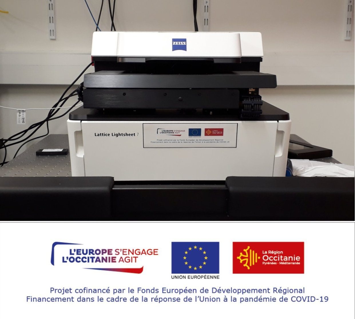
Tarif établissement partenaire: -- €/heure
Tarif établissement public et privé : -- €/heure
Objectives :
10X Plan Apochromat 0.45 NA
20X Plan Apochromat 0.8 NA
40X Plan Apochromat 1.3 NA oil DIC
63X Plan Apochromat 1.4NA oil DIC
Détecteur :
1 caméra Leica K5 sCMOS monochrome
(2048 x 2048 pixels, 6.5µm taille du pixel)
Environnement L1
Light sources :
LED3 Leica for episcopic illumination
White LED for diascopic illumination
Motorization:
Binocular tube, stage (X, Y and Z), fluorescence filter set, condenser and ,objective turrets.
Fluorescence filter sets:
DAPI Exc 405/60, FT465, Em470/40
GFP HE Exc 470/40, FT495, Em 525/50
Cy3 Exc 545/30, FT570, Em 610/75
Cy5 Exc 620/60, FT660, Em 700/75
Available acquisition techniques:
Any slide acquisition automation in both fluorescence and transmission modes.
Mosaic acquisition (full slide scan, slices on a slide scan) with user defined autofocus map.
Personnalisation des protocoles.
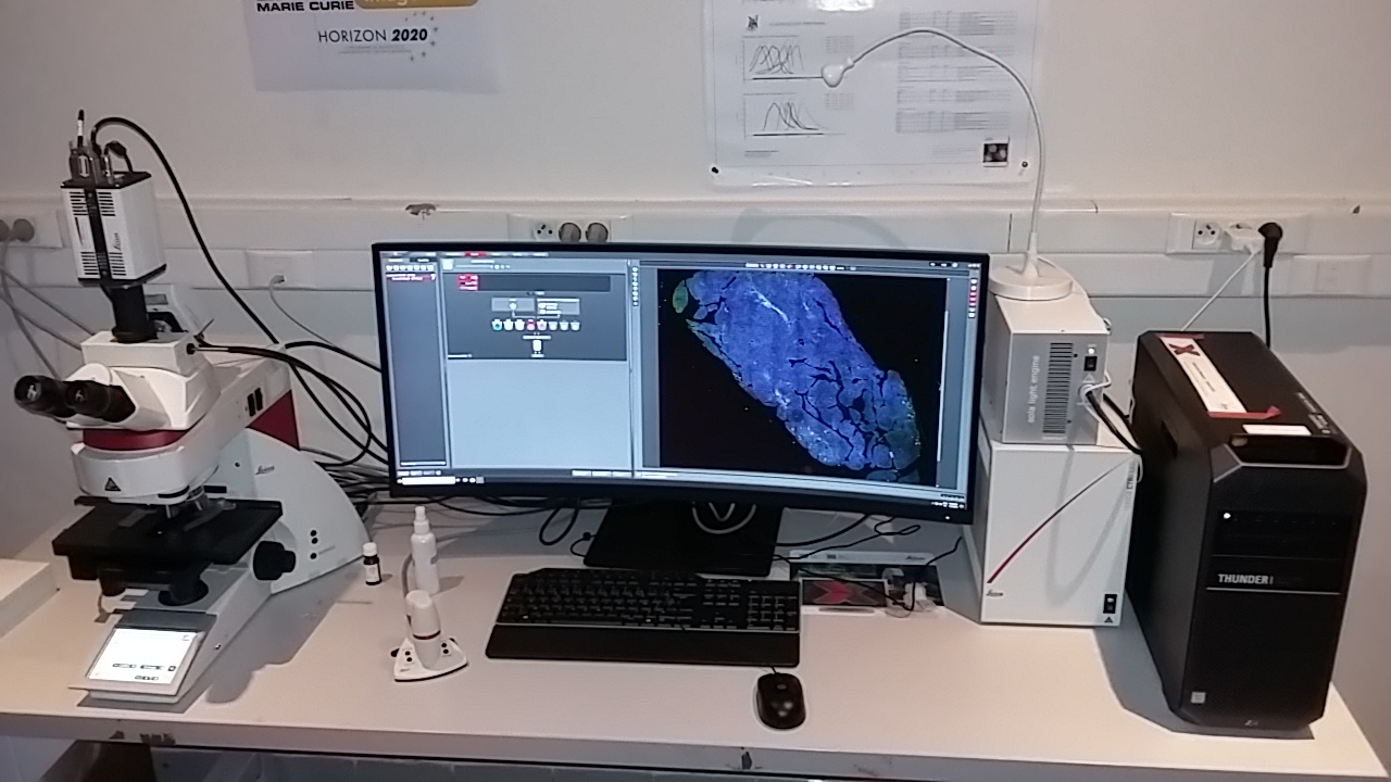
Image quality :
Quality and resolution improvement in real time or post processing mode with Leica THUNDER technology :
Instant Computational Clearing (ICC in 2D)
Large Volume Computational Clearing (LVCC in 3D)
Inquiries may be sent to This email address is being protected from spambots. You need JavaScript enabled to view it.
Posted in Widefield microscopy
Partner lab price : 37.04 €/hour
Public and private lab price: 101.89 €/hour
Objectives:
10X Plan Apochromat 0.4 NA Imm
20X Plan Apochromat 0.75 NA Imm Corr
40X Plan Apochromat 1.3 NA oil DIC
63X Plan Apochromat 1.4 NA oil DIC
63X Plan Apochromat 1.2W UVIS + water dispenser
Detectors:
2 cooled regular multialkaline PMTs
1 transmission PMT
3 cooled HyDs (GaAsP photocathode) for low noise photon counting imaging.
BSL1 Environment
Illumination light source for widefield observation :
Fibred metal halide arc source for episcopic illumination
100W tungsten bulb for diascopic illumination
Motorization:
Binocular tube, X, Y and Z stage, fluorescence filter set turret, transmission mirror, objective turret
Fluorescence filter sets for widefield observation:
Hoechst
GFP
Rhodamine LP
Cy5
Available acquisition techniques:
Excitation with 355, 405, 458, 476, 488, 514, 561 & 633nm lines.
2/3D mosaic acquisition, spectral separation, timelapse analysis. In-focus repositioning.
Any photoactivation/destruction technique (FRAP, PA, FLIP), ratiometric FRET experiment, UVA induced DNA-damange on sensitized cells.
Screening and on the fly analysis (Matrix screener)
User-defined protocols
Image quality:
Confocal technology allows improved spatial resolution and contrast compared to widefield imaging. Spatial resolution may be further improved using deconvolution algorithms (Huygens).
Sensitivity:
The HyD design allows better sensitivity and SNR compared to regular PMT and a less noisy photon counting imaging.
The fast resonant scanner decrease excitation-related toxicity.
Inquiries may be sent to This email address is being protected from spambots. You need JavaScript enabled to view it.
Posted in Confocal microscopy
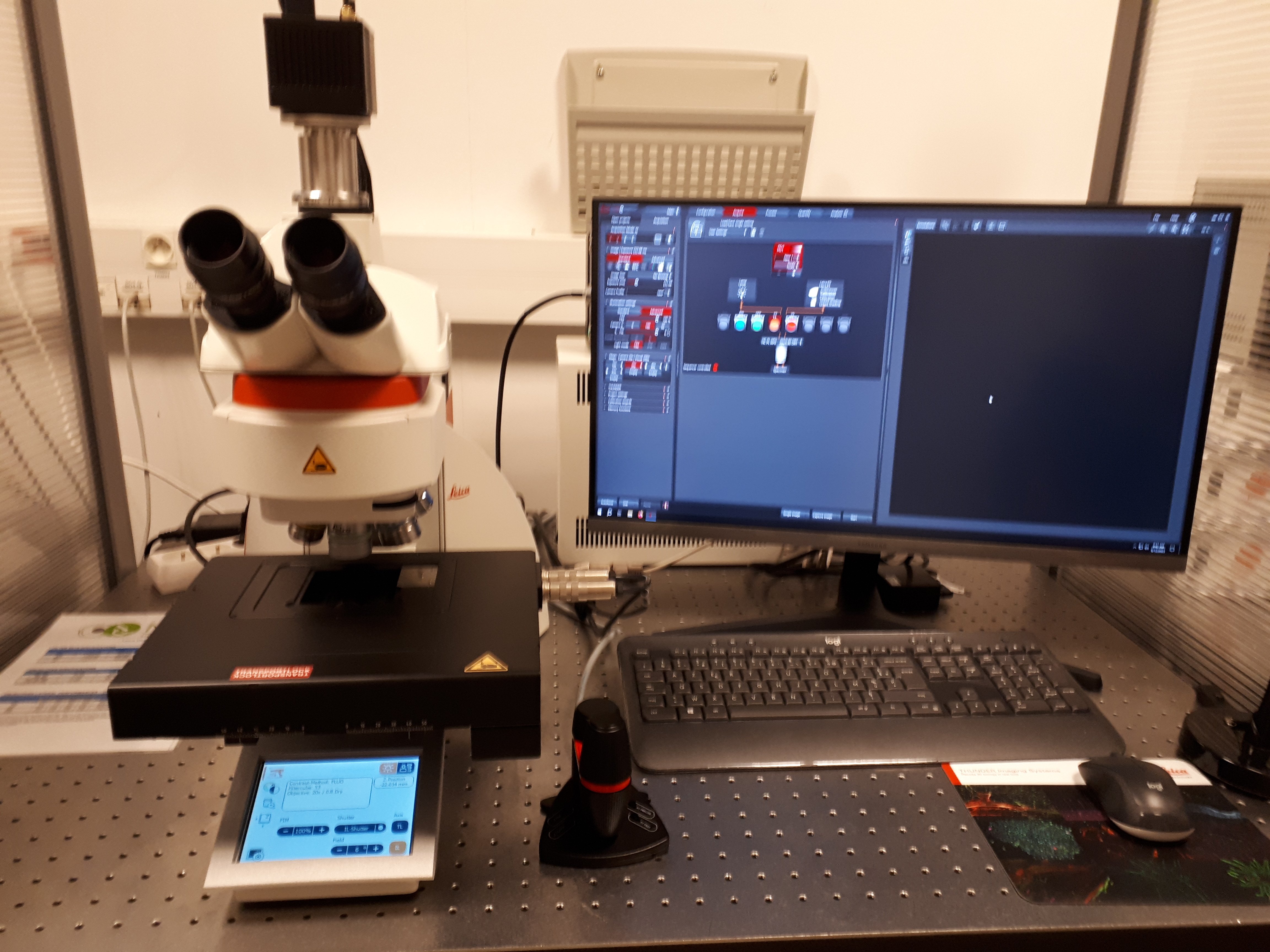
Tarif établissement partenaire: 15.66 €/heure
Tarif établissement public et privé : 55.41 €/heure
1.25X/0.04 HC PL Fluotar (Working Distance=3.7mm)
10X/0.32 HC PL Fluotar (WD=11.13mm)
20X/0.55 HC PL Fluotar (WD=1.2mm)
40X/1.3 Oil HC PL APO DIC (WD=0.22mm)
63X/1.4 Oil HC PL APO DIC (WD=0.14mm)
100X/1.4 Oil HC PL APO DIC Ph (WD=0.09mm)
Illumination source LED for Transmission and Fluo (SOLA)
filter cubes :
DAPI Ex360/40, FT400, Em470/40
GFP Ex470/40, FT495, Em525/50
RHOD Ex546/10, FT560, Em585/40
TxRed Ex560/40, FT585, Em630/75
Cy5 Ex620/60, FT660, Em700/75
Color camera for histology :
CMOS 3.1MP (2048x1536, pixel size = 3.2µm)
B&W camera for fluorescence :
K8 sCMOS 4.2MP (2048x2048, pixel size = 6.5µm)
software : LAS X
Multidimentional acquisition : 2D, 3D+time multicolor
Navigator Module for fast and easy mosaic, multiposition and focus correction
quality and resolution improvement in real time or post processing with Leica THUNDER technology :
- Instant Computational Clearing (ICC in 2D) = real time identification of out of focus signal, debluring
- Large Volume Computational Clearing (LVCC in 3D) = ICC + adaptative deconvolution
preserve raw data, quantifiable, adapted for thick and photosensitive samples
Posted in Widefield microscopy
")
")