")
")
English
Compensations
If you use several fluorochromes (for example A and B) in a single experiment, the emission spectra may overlapped. Thus, it is necessary to compensate the signals, that is to say to eliminate the fluorescence provided by the A fluorochrome in the channel where you read the B fluorescence.
To well set up the compensations, you need mono-staining of each used fluorochrome in the experiment.
Please, contact the manager of the facility to optimize your fluorochrome choices regarding the cytometer installed on the facility.
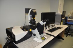
Confocal Leica SP8
multidimensional scan mode
3D imaging on fixed or live cell sample
suitable for F-techniques (FRAP - FRET)
Partner lab price : 37.04 €/hour
Public and private lab price: 101.89 €/hour
Brief description:
motorized inverted confocal microscopemultidimensional scan mode
3D imaging on fixed or live cell sample
suitable for F-techniques (FRAP - FRET)
10X HC Plan Fluotar 0.3 NA
20X HC Plan Apochromat 0.7 NA CS
40X HCX corr Plan Apochromat CS 1.1 NA water
40X HCX Plan Apochromat CS 1.3 NA oil
63X HCX Plan Apochromat CS 1.4 NA oil
20X HC Plan Apochromat 0.7 NA CS
40X HCX corr Plan Apochromat CS 1.1 NA water
40X HCX Plan Apochromat CS 1.3 NA oil
63X HCX Plan Apochromat CS 1.4 NA oil
exciation source : Lamp HXP120
Lasers : 405, Argon (458, 488, 514), 561, 633
Filters cubes
DAPI (Ex BP340-380, Em LP425
FITC (Ex Bp450-490, Em LP515
GFP (Ex BP480/40, Em BP527/30)
DsRed Et,k (Ex BP546/12, Em BP605/75)
spectral acquisition in confocal mode
Lasers : 405, Argon (458, 488, 514), 561, 633
Filters cubes
DAPI (Ex BP340-380, Em LP425
FITC (Ex Bp450-490, Em LP515
GFP (Ex BP480/40, Em BP527/30)
DsRed Et,k (Ex BP546/12, Em BP605/75)
spectral acquisition in confocal mode
Détectors : 1 PMT and 2 PMTs hybride HyD GaAsP
multi-dimensional scan mode: XYZ, XZ etc... lambda, time lapse, mark and find, tile scan
FRAP / FRET
FRAP / FRET
room working temperature between 19 to 26°C
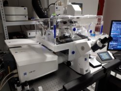
confocal LSM880 Airyscan
Confocal Zeiss LSM880 Airyscan
+ DIC images
Controled by Zen 2012 software
Partner lab price : 37.04 €/hour
Public and private lab price: 101.89 €/hour
Brief description:
Inverted confocal microscope for 3D imaging of fixed sample (slide-coverslip) or live sample (bottom glass coverslip dish). Improvment of spatial resolution thank to the Airyscan module and speed of acquisition thank to the Fast Airyscan option+ DIC images
Controled by Zen 2012 software
Objectives:
10X EC Plan Neofluar 0.3NA
20X Plan Apo 0.8NA
40X Plan Apo huile 1.3NA
63X Plan Apo huile 1.4NA
10X EC Plan Neofluar 0.3NA
20X Plan Apo 0.8NA
40X Plan Apo huile 1.3NA
63X Plan Apo huile 1.4NA
Illumination source for occular widefield visualisation:
HXP120 lamp
Cubes for occular widefield visualisation: DAPI, GFP, Cy3
Excitation sources for confocal mode:
Diode 405nm
Argon LASER (458, 488, 514nm)
DSSP 561nm
Helium/Neon LASER 633nm
HXP120 lamp
Cubes for occular widefield visualisation: DAPI, GFP, Cy3
Excitation sources for confocal mode:
Diode 405nm
Argon LASER (458, 488, 514nm)
DSSP 561nm
Helium/Neon LASER 633nm
Detectors:
-2 PMT for fluorescence
-1 internal GaAsP detector
-1 PMT for transmission
-1 Airyscan detector (32 GaAsP detectors)
Environment control: temperature and CO2
-2 PMT for fluorescence
-1 internal GaAsP detector
-1 PMT for transmission
-1 Airyscan detector (32 GaAsP detectors)
Environment control: temperature and CO2
Acquisition mode:
Zeiss Zen Software
-multi-modal acquisition: XYZ, spectral, time, multi-positions or mosaic
-"super-resolution" imaging
-hardware auto-focus Definite Focus
-FRAP module
-3D mosaic module
Zeiss Zen Software
-multi-modal acquisition: XYZ, spectral, time, multi-positions or mosaic
-"super-resolution" imaging
-hardware auto-focus Definite Focus
-FRAP module
-3D mosaic module
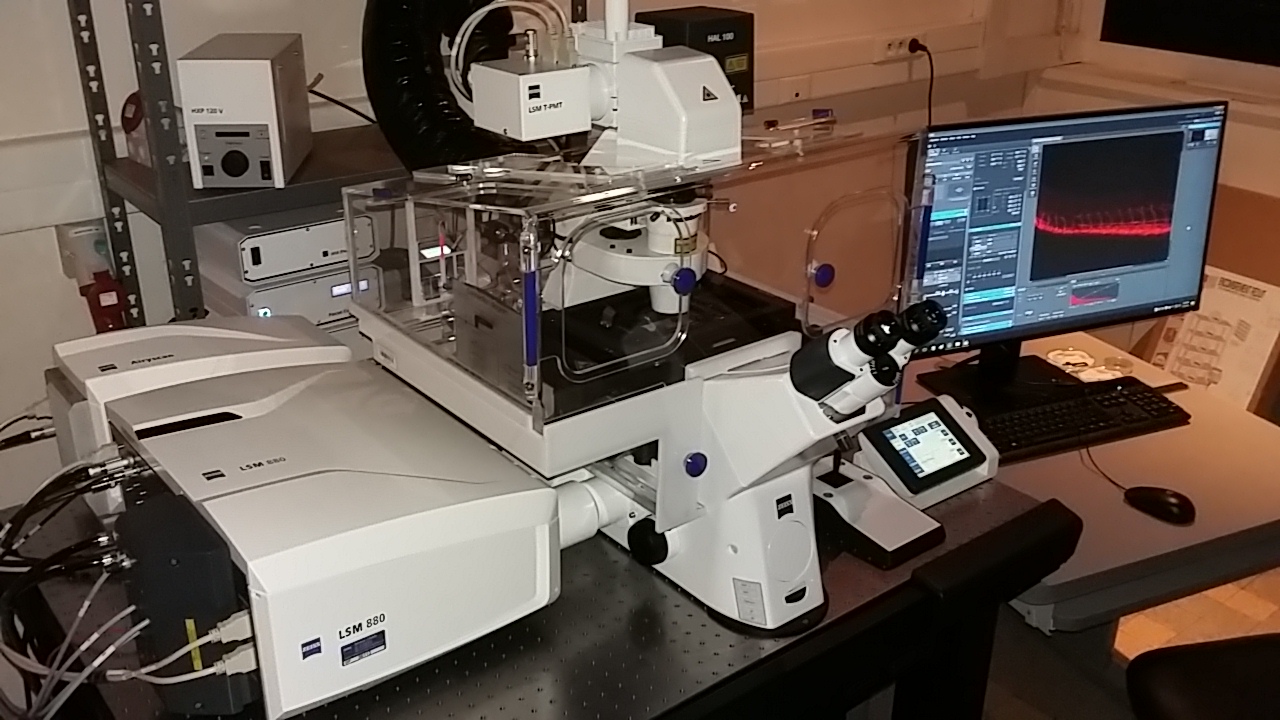
Confocal ZEISS LSM880 FastAiryscan
Imagerie 3D
Vidéomicroscopie
Super-résolution
FRAP photoactivation
Imagerie in vivo
Imagerie spectrale
Improvement of spatial resolution and speed with Airyscan and FastAiryscan module.
Microscope equiped with DIC and autofocus
Tarif établissement partenaire: 37.04 €/heure
Tarif établissement public et privé : 101.89 €/heure
Technologies associées :
Brève description :
Inverted confocal microscope for 3D imaging on fixed (slide/coverslip) or live samples (bottom glass petridish). Improvement of spatial resolution and speed with Airyscan and FastAiryscan module.
Microscope equiped with DIC and autofocus
Objectives :
10X/0.3 Plan-neofluar
20X/0.8 Plan-apochromat
40X/1.1 W C-apochromat (long distance) DIC
40X/1.4 Oil Plan-apochromat DIC
63X/1.4 Oil Plan-apochromat DIC
Occular fluorescence with HXP120
filter cubes : DAPI, GFP, Cy3
10X/0.3 Plan-neofluar
20X/0.8 Plan-apochromat
40X/1.1 W C-apochromat (long distance) DIC
40X/1.4 Oil Plan-apochromat DIC
63X/1.4 Oil Plan-apochromat DIC
Occular fluorescence with HXP120
filter cubes : DAPI, GFP, Cy3
Lasers :
Diode 405
Laser Argon 458, 488, 514
DSSP 561
Laser He/Ne 633
Detectors :
- 2 PMT fluorescence
- 1 GaAsP (high sensitivity)
- 1 PMT Transmission
- 1 Airyscan detector (32 GaAsP)
Acquisition software : ZEN black (.czi files format)
Diode 405
Laser Argon 458, 488, 514
DSSP 561
Laser He/Ne 633
Detectors :
- 2 PMT fluorescence
- 1 GaAsP (high sensitivity)
- 1 PMT Transmission
- 1 Airyscan detector (32 GaAsP)
Acquisition software : ZEN black (.czi files format)
- multi modal aquisitions : XYZ, time, spectral, multiposition, mosaic
- "super-resolution" imaging with Airyscan and FastAiryscan module
- hardware solution for autofocus (Definite focus)
- FRAP, FRET, Photoactivation module
- Experiment designer module
- "super-resolution" imaging with Airyscan and FastAiryscan module
- hardware solution for autofocus (Definite focus)
- FRAP, FRET, Photoactivation module
- Experiment designer module
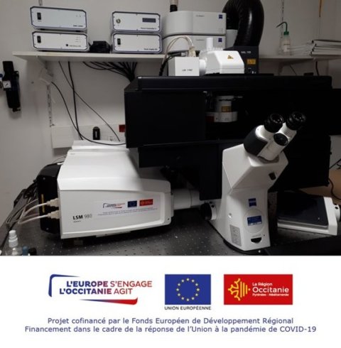
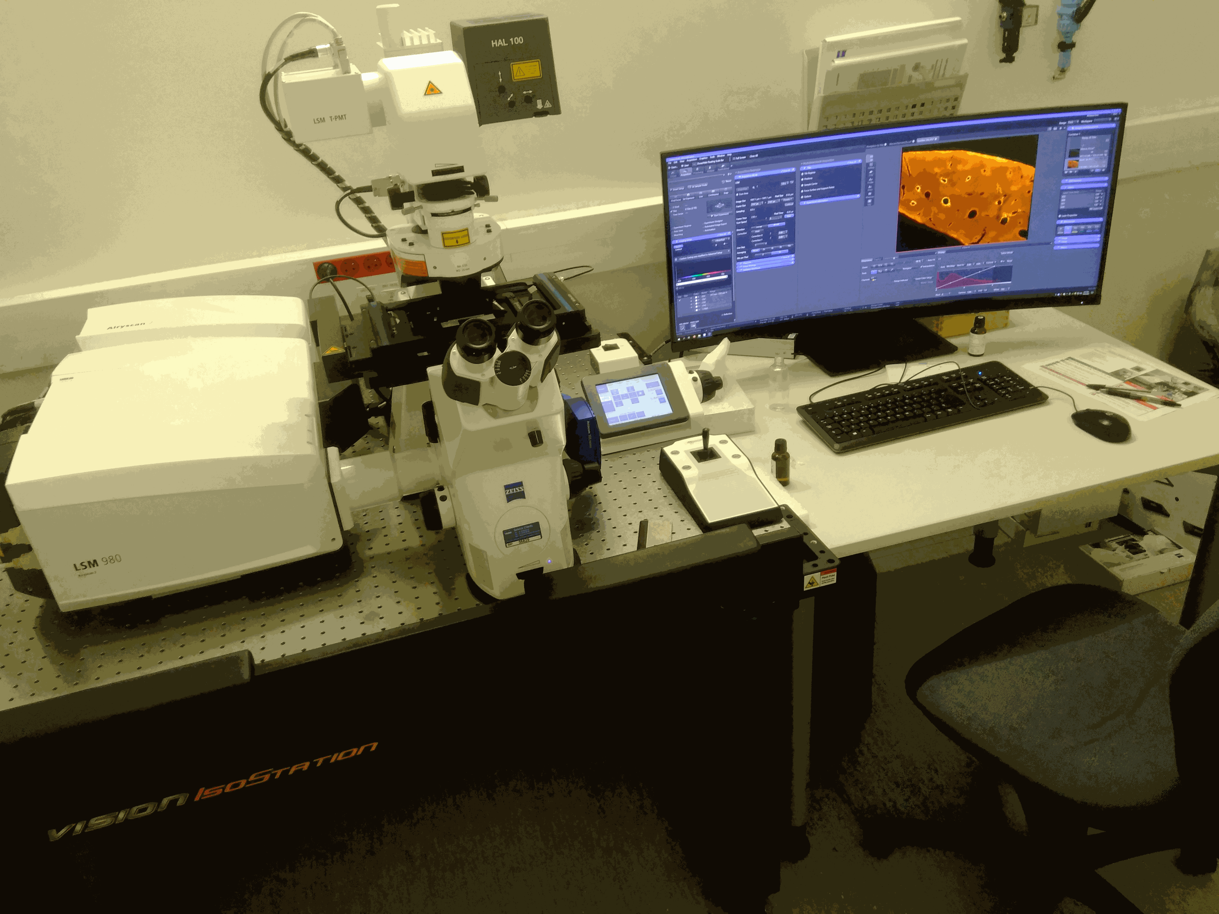
CONFOCAL ZEISS LSM980 AIRYSCAN
Tarif établissement partenaire: 37.04 €/heure
Tarif établissement public et privé : 101.89 €/heure
Brève description :
Inverted confocal microscope for 3D imaging of fixed sample (slide-coverslip) or live sample (bottom glass coverslip dish). Improvment of spatial resolution thank to the Airyscan 2 module and speed of acquisition thank to the AiryScan Multiplex 4Y & 8Y.
Objectives:
20X Plan Apo 0.8NA
40X Plan Apo huile 1.3NA
63X Plan Apo huile 1.4NA
40X Plan Apo eau 1.1NA
20X Plan Apo 0.8NA
40X Plan Apo huile 1.3NA
63X Plan Apo huile 1.4NA
40X Plan Apo eau 1.1NA
Detectors:
-2 PMT for fluorescence
-1 GaAsP spectral detector 32 chanels
-1 PMT for transmited light
-1 AiryScan2 detector
-2 PMT for fluorescence
-1 GaAsP spectral detector 32 chanels
-1 PMT for transmited light
-1 AiryScan2 detector
Illumination source for ocular widefield visualisation :
Colibri 5
Cube for widefield visualisation : 3 bands DAPI/GFP/CY3
Environment control: Temperature, CO2 and humidity
Excitation sources for confocal mode :
Laser diodes 405nm, 445nm, 488nm, 515nm, 561nm and 639nm
Colibri 5
Cube for widefield visualisation : 3 bands DAPI/GFP/CY3
Environment control: Temperature, CO2 and humidity
Excitation sources for confocal mode :
Laser diodes 405nm, 445nm, 488nm, 515nm, 561nm and 639nm
Acquisition modes:
Zeiss Zen Blue Software
-multi-modal acquisition: XYZ, spectral, time, multi-positions & mosaic
-« super-resolution » imaging for fixed samples
-Live and « super-resolution » imaging with the multiplex 4Y & 8Y AiryScan
-AI Sample Finder module
-FRAP module
Zeiss Zen Blue Software
-multi-modal acquisition: XYZ, spectral, time, multi-positions & mosaic
-« super-resolution » imaging for fixed samples
-Live and « super-resolution » imaging with the multiplex 4Y & 8Y AiryScan
-AI Sample Finder module
-FRAP module
Confocal Zeiss LSM980 Airyscan 8Y
3D imaging
High resolution
FRAP photoactivation
Spectral imaging
Image analysis
Controled by Zen Blue.
Partner lab price : 37.04 €/hour
Public and private lab price: 101.89 €/hour
Associated technologies:
Brief description:
Inverted confocal microscope for 3D imaging of fixed sample (slide-coverslip) or live sample (bottom glass coverslip dish). Improvment of spatial resolution thank to the Airyscan 2 module and speed of acquisition thank to the AiryScan Multiplex 4Y & 8Y.Controled by Zen Blue.
Objectives:
5X Plan Neofluar 0.16NA
10X Plan Apo 0.45NA
20X Plan Apo 0.8NA
40X Plan Apo oil 1.3NA
63X Plan Apo oil 1.4NA
5X Plan Neofluar 0.16NA
10X Plan Apo 0.45NA
20X Plan Apo 0.8NA
40X Plan Apo oil 1.3NA
63X Plan Apo oil 1.4NA
Illumination source for ocular widefield visualisation :
Colibri 7
Cubes for ocular widefield visualisation : 3 bandes DAPI/GFP/CY3
Excitation sources for confocal mode :
Diode 405nm
Diode 488nm
Diode 561nm
Diode 639nm
Colibri 7
Cubes for ocular widefield visualisation : 3 bandes DAPI/GFP/CY3
Excitation sources for confocal mode :
Diode 405nm
Diode 488nm
Diode 561nm
Diode 639nm
Detectors:
-2 PMT for fluorescence
-1 internal GaAsP detector
-1 PMT for transmission
-1 Airyscan 2 detector
Environment control : no
Environment control: not available
-2 PMT for fluorescence
-1 internal GaAsP detector
-1 PMT for transmission
-1 Airyscan 2 detector
Environment control : no
Environment control: not available
Acquisition mode:
Zeiss Zen Blue Software
-multi-modal acquisition: XYZ, spectral, time, multi-positions & mosaic
-« super-resolution » imaging for fixed samples
-Live and « super-resolution » imaging with the multiplex 4Y & 8Y AiryScan
-AI Sample Finder module
-FRAP module
Zeiss Zen Blue Software
-multi-modal acquisition: XYZ, spectral, time, multi-positions & mosaic
-« super-resolution » imaging for fixed samples
-Live and « super-resolution » imaging with the multiplex 4Y & 8Y AiryScan
-AI Sample Finder module
-FRAP module
Confocal Zeiss LSM980 NLO
3D imaging
FRAP photoactivation
FCS / FCCS
FRET / FLIM
SHG / THG
Spectral imaging
Partner lab price : 37.04 €/hour
Public and private lab price: 101.89 €/hour
Associated technologies:
Brief description:
Inverted confocal microscope coupled with a multiphoton laser for 3D imaging of fixed sample (slide-coverslip) or live sample (bottom glass coverslip dish) and for F-technics (FRAP, photactivation, FCS, FCCS, FLIM) and SHG detection.
Objectives:
10X EC Plan Neofluar 0.3NA
20X Plan Apo 0.8NA
40X Plan Apo oil 1.3NA
63X Plan Apo oil 1.4NA
40X C-APO water 1.4NA
10X EC Plan Neofluar 0.3NA
20X Plan Apo 0.8NA
40X Plan Apo oil 1.3NA
63X Plan Apo oil 1.4NA
40X C-APO water 1.4NA
Illumination source for occular widefield visualisation:
HXP120 lamp
Cubes for occular widefield visualisation: DAPI, GFP, Cy3
Sources d'excitation mode confocal :
Diode 405nm
Diode 488nm
Diode 561nm
Diode 639nm
Multiphoton laser:
Chameleon Ultra-II Coherent LASER (680-1080nm)
HXP120 lamp
Cubes for occular widefield visualisation: DAPI, GFP, Cy3
Sources d'excitation mode confocal :
Diode 405nm
Diode 488nm
Diode 561nm
Diode 639nm
Multiphoton laser:
Chameleon Ultra-II Coherent LASER (680-1080nm)
Detectors:
-2 PMT fluorescence
-1 multi-PMT (32 détecteurs GaAsP) spectral detectorl
-1 PMT transmission
-1 camera for tilescan preview
-1 detector FLIM (photon counting for FLIM of GFP or Cy3)
Temperature and CO2 control
-2 PMT fluorescence
-1 multi-PMT (32 détecteurs GaAsP) spectral detectorl
-1 PMT transmission
-1 camera for tilescan preview
-1 detector FLIM (photon counting for FLIM of GFP or Cy3)
Temperature and CO2 control
Acquisition mode:
Zeiss Zen Blue Software
-multi-modal acquisition: XYZ, spectral, time, multi-positions & mosaic
-FRAP module
Software Becker Hickl SPCM et SPC-100
-module FLIM (cart and detector Becker-Hickl)
Zeiss Zen Blue Software
-multi-modal acquisition: XYZ, spectral, time, multi-positions & mosaic
-FRAP module
Software Becker Hickl SPCM et SPC-100
-module FLIM (cart and detector Becker-Hickl)
Contact us
About an order or a problem in the subscription process
Send an e-mail to à This email address is being protected from spambots. You need JavaScript enabled to view it.
To contact a specific MRI facility
As a general rule, contact the person in charge of the targeted facility, whose contact details can be found on the webpage dedicated to the facility and in the directory.
Alternatively, you can use the generic addresses of facilities with several engineers:
Send an e-mail to This email address is being protected from spambots. You need JavaScript enabled to view it. to contact MRI imaging facility located at the CRBM
Send an e-mail to This email address is being protected from spambots. You need JavaScript enabled to view it. to contact MRI imaging facility located at the IGH
Send an e-mail to This email address is being protected from spambots. You need JavaScript enabled to view it. to contact MRI imaging facility located at the INM
Send an e-mail to This email address is being protected from spambots. You need JavaScript enabled to view it. to contact MRI flow cytometry facility located at the IGMM
Send an e-mail to This email address is being protected from spambots. You need JavaScript enabled to view it. to contact MRI flow cytometry facility located at the IRMB
Send an e-mail to This email address is being protected from spambots. You need JavaScript enabled to view it. to contact MRI image analysis service MRI-CIA
For any other subject or if you don't know who to contact :
Send an e-mail to This email address is being protected from spambots. You need JavaScript enabled to view it.